Purpose
Sonus Plus system is intended to perform ultrasound studies of internal and surface organs. It is suitable for use in hospital, outpatient departments, as well as in private medical and veterinary practice.
Studies in all areas of ultrasound diagnostics:
- clinic of internal diseases;
- surgery;
- gynecology;
- obstetrics;
- urology;
- pediatrics;
- neonatology;
- cardiology;
- endocrinology;
- angiology;
- gastroenterology;
- neurology;
- orthopedics;
- transcranial studies.
Specification
Technologies of scanning and image processing:
- anatomic M-mode;
- estimation of elasticity of blood vessels;
- color mapping of pulsating blood flow;
- automatic evaluation of the intima-media thickness;
- compression elastography;
- assessment of the attenuation coefficient of the tissue;
- trapezoid imaging for linear probe;
- improved image quality and suppression of speckle noise;
- multiangle compound imaging;
- tissue Doppler;
- inverse harmonic;
- sector scanning;
- synthetic aperture;
- vector scanning.
Specifications:
- 2 ports for probes connection;
- possibility to increase the number of probes to be connected up to 4 pcs. via port extender (optional);
- weight no more than 14.0 kg;
- time of continuous operation 12 hours;
- total dynamic range 263 dB;
- frequency range from 1 MHz to 14 (broadband system);
- 7 zones gain adjustment in depth;
- control of the power level of acoustic signal;
- up to 8 simultaneous focuses;
- dynamic focusing;
- dynamic aperture;
- dynamic apodization;
- adaptive image processing filter;
- linear image magnification up to 16 times;
- automatic optimization of the echo images in B-, D-, as well as duplex and triplex modes;
- conducting specialized measurement with results to be entered into the protocol;
- multifrequency (more than three frequencies for each probe), with the user's ability to choose how to control the frequency — coarse and fine;
- evenly depth control step;
- quick regulation of time gain control slope;
- maintaining of patients data base;
- ultrasound images archiving system to the internal disk with the capacity of not less than 1 TB;
- protocols and conclusions templates;
- availability of factory default settings of scanning and processing parameters for each type of study and probe;
- ability to create custom settings for each type of study and probe;
- support up to 5 users;
- echo images and cineloops postprocessing;
- record of study's results on the USB flash drive;
- image gallery;
- ability to perform puncture and biopsy;
- saving and extradition of images in DICOM standard.
Imaging modes:
- B, B/B, 4B – two-dimensional gray-scale;
- THІ – B (M) modes on tissue (second) harmonic;
- Inv H – B (M) modes on the inverse harmonic;
- M (B/M) – one-dimensional (combined two-dimensional and one-dimensional) gray-scale scan mode with time-base sweep;
- MA – anatomic mode;
- CD (B/CD) – color mapping of blood flow velocity;
- PD (B/PD) – the color mapping of energy (energy and direction) of the Doppler signal;
- DF (B/DF) – color mapping of dynamic flow;
- color mapping of pulsatile blood flow (VP);
- CD(PD, DF, TD)/CD(PD, DF, TD) – alternately modes on two windows;
- 4CD(PD, DF, TD) – modes in a circle on 4 window;
- MC(MCD, MPD, MDF, MTD) – color mapping of M-mode;
- CD(PD, DF, TD)/MCD(PD, DF, TD) – color mapping of B- and M-modes;
- D(PW) – PW spectral Doppler with all probes;
- B/D – PW duplex and alternately duplex;
- CD(PD, DF, TD)/D(PW) – triplex and alternately triplex;
- E-AS – estimation of elasticity of blood vessels;
- E-IMT – automatic evaluation of intima-media complex thickness.
Probes
|
 C205E C205E
(50R; 1,5-5,0 MHz; 128 el)
|
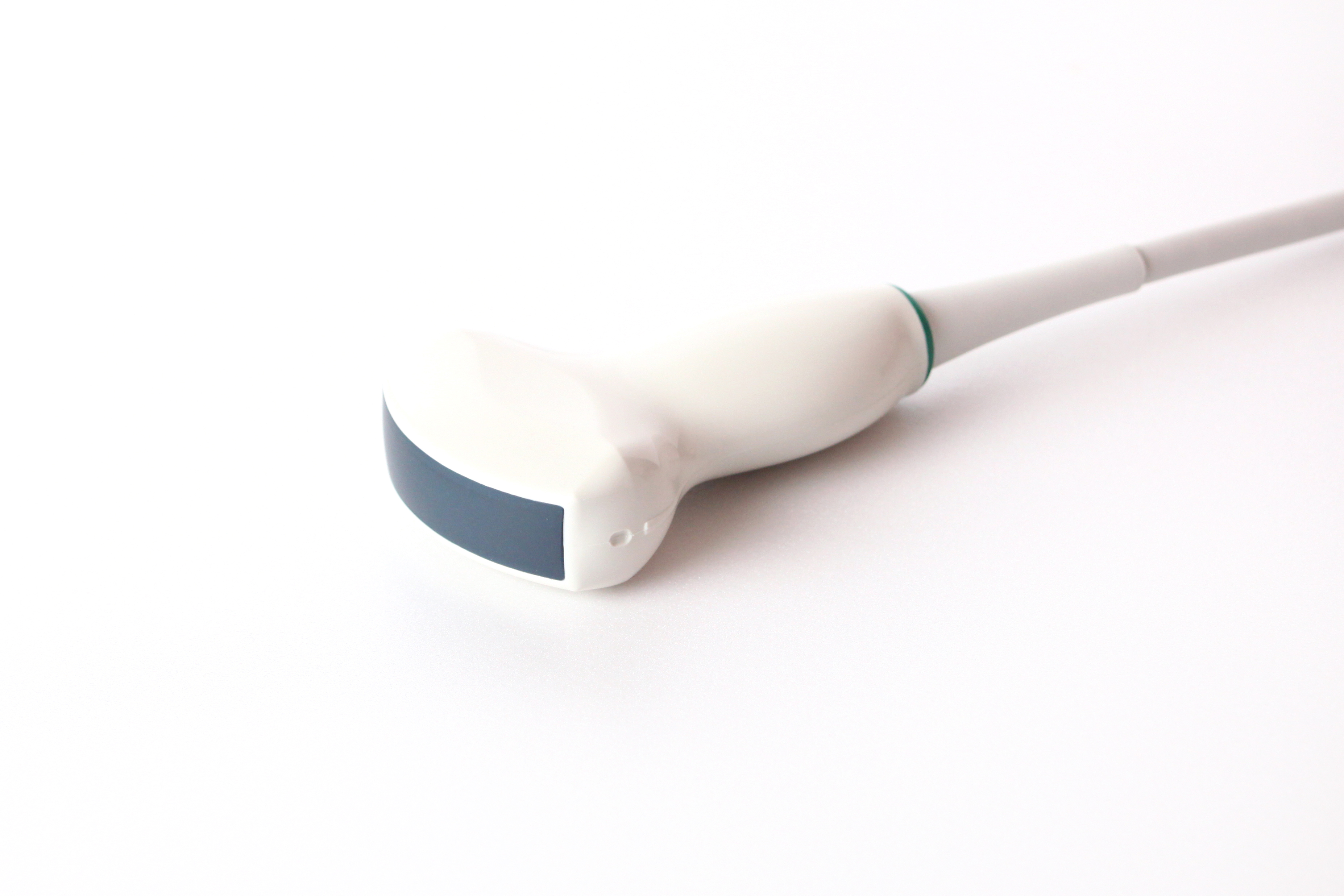 C1-5/60EP C1-5/60EP
(60R; 1,5-5,0 MHz; 128 el)
|
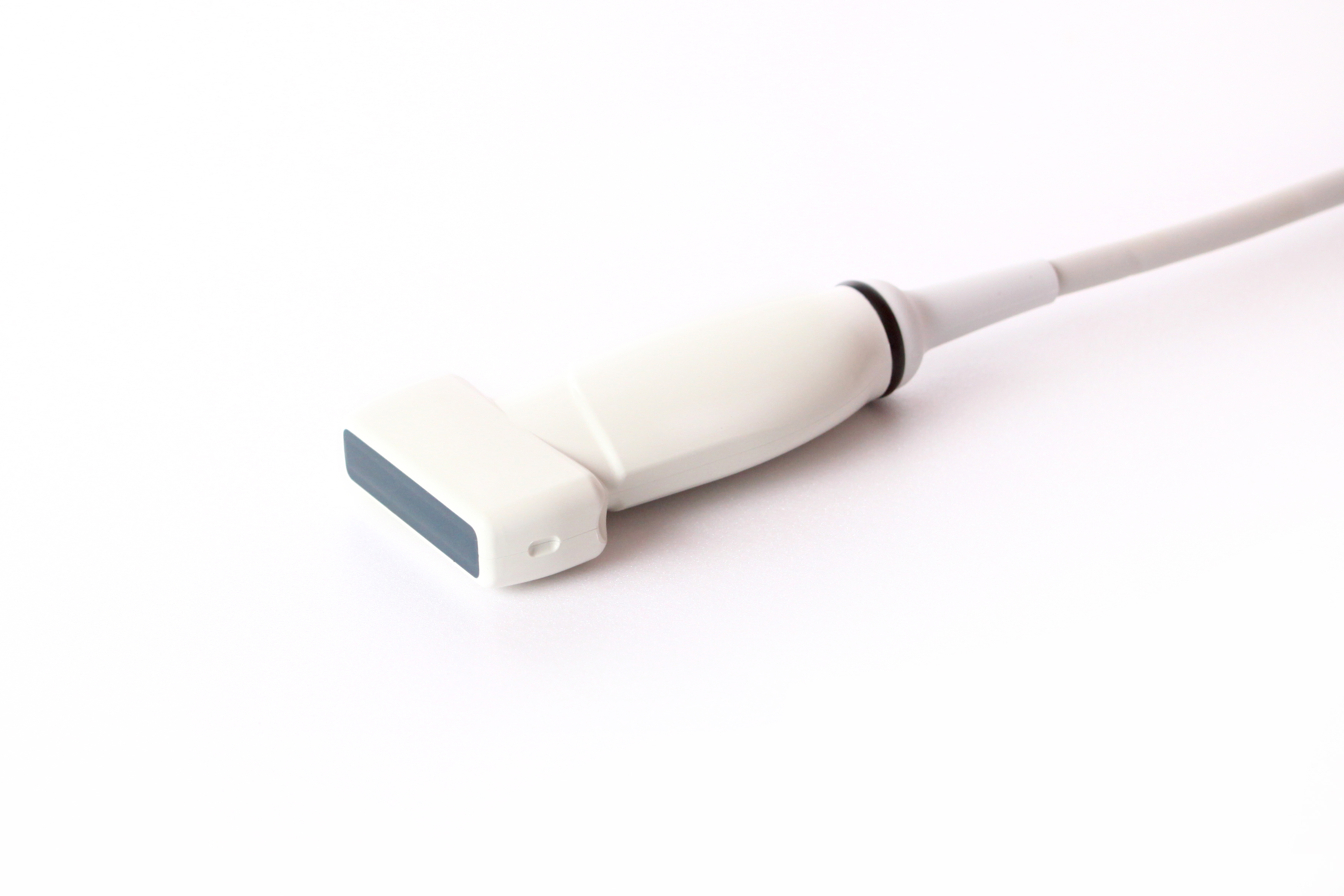 L224E L224E
(38 mm; 4,5-10,0 MHz; 128 el)
|
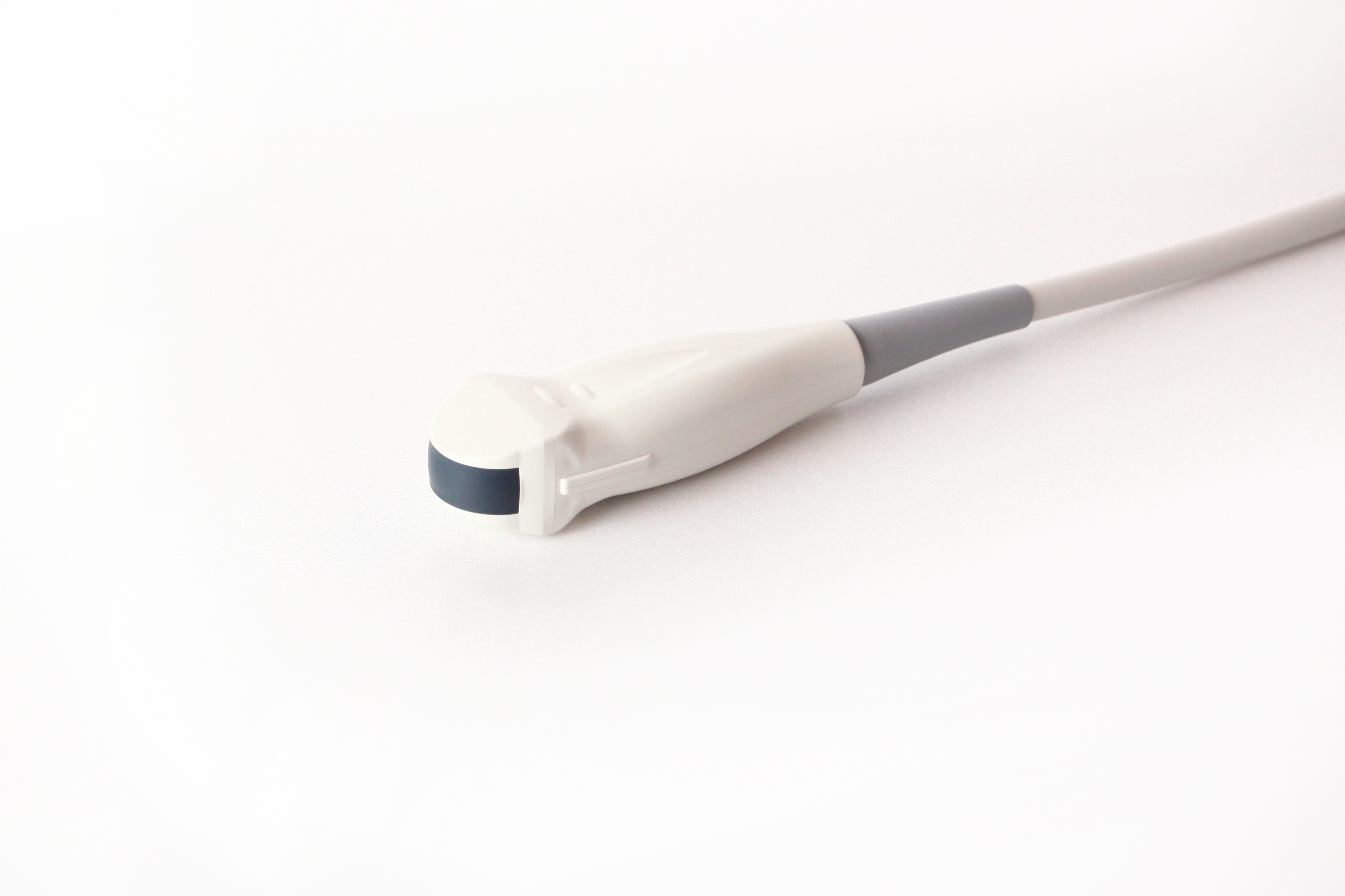 MC231E MC231E
(11,5R; 3,60-9,0 MHz; 128 el)
|
|
 EC231E EC231E
(11,5R; 3,6-9,0 MHz; 128 el)
|
|
|
|
|
We recommend using the contact gel for ultrasound examinations TU U 16475627.003-96 of Eskulap-UST Company. The use of the gel from other manufacturers could cause probe's damage.
|
Gallery
This section is under construction.
Brochure
This section is under construction.
Certificates
- Certificate of Conformity (with Annex) – Browse
- Certificate of Quality Management System – Browse

